Key takeaways
- A research lab doing routine cell culture checks and basic histology work needs a different microscope than one running fluorescence-based imaging or advanced live-cell studies, so the right pick depends mostly on illumination type, objective quality, and whether fluorescence or…
A research lab doing routine cell culture checks and basic histology work needs a different microscope than one running fluorescence-based imaging or advanced live-cell studies, so the right pick depends mostly on illumination type, objective quality, and whether fluorescence or phase contrast capability is actually required for the work planned. Compound light microscopes with good optics and Kohler illumination cover most routine biological research; fluorescence-capable or advanced research-grade scopes are only worth the jump for labs that specifically need those imaging modes.
As an Amazon Associate, we earn from qualifying purchases made through links on this page.
Compound light microscopes: the research workhorse
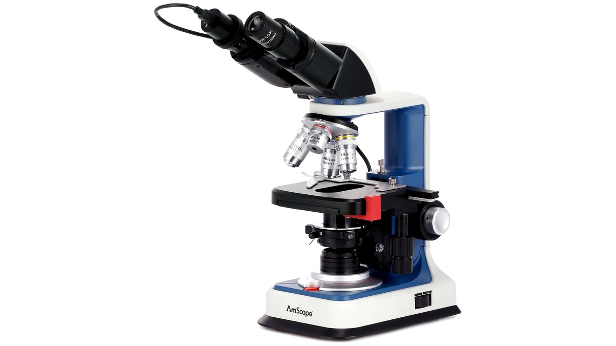
Most biological research, from student labs to working research groups, is built around a compound light microscope using transmitted brightfield illumination, typically with a set of objectives covering low (4x or 10x), intermediate (20x or 40x), and high (60x or 100x, often oil-immersion) magnification. Instrument makers and microscopy outlets consistently stress that objective and condenser quality matter more to actual image sharpness and resolution than magnification numbers alone, since a cheap high-magnification objective can produce a dimmer, blurrier image than a well-made lower-magnification one. Proper Kohler illumination, which evenly lights the specimen and allows adjustable aperture and field diaphragms, is a feature researchers and microscopy specialists consistently recommend checking for on any scope intended for serious lab use rather than casual viewing.
Phase contrast and other contrast-enhancing techniques
Many biological samples, particularly live, unstained cells, are nearly transparent under standard brightfield illumination, which is why labs doing cell culture work commonly add phase contrast optics, a technique that converts subtle differences in refractive index within the specimen into visible contrast without staining. Darkfield illumination is another contrast option some labs use for specific specimen types, producing a bright image against a dark background that can reveal edges and fine structures standard brightfield misses. Both techniques typically require specific objectives and condenser components matched to the technique, so a scope bought for brightfield only cannot simply add phase contrast later without the matched hardware.
Fluorescence capability: a significant step up
Labs doing immunofluorescence, studying tagged proteins, or working with fluorescent reporter constructs need a fluorescence microscope, which adds a specific light source (commonly an LED, mercury, or metal-halide fluorescence illuminator depending on the model and era), filter cubes matched to the fluorophores in use, and objectives optimized for fluorescence work. This is a genuinely different and more expensive category than a standard compound scope, and makers and microscopy core facilities generally advise researchers to confirm which specific fluorophores or filter sets they’ll need before buying, since filter cubes are usually sold to match particular excitation and emission wavelength ranges rather than being universal.
Camera integration and digital imaging
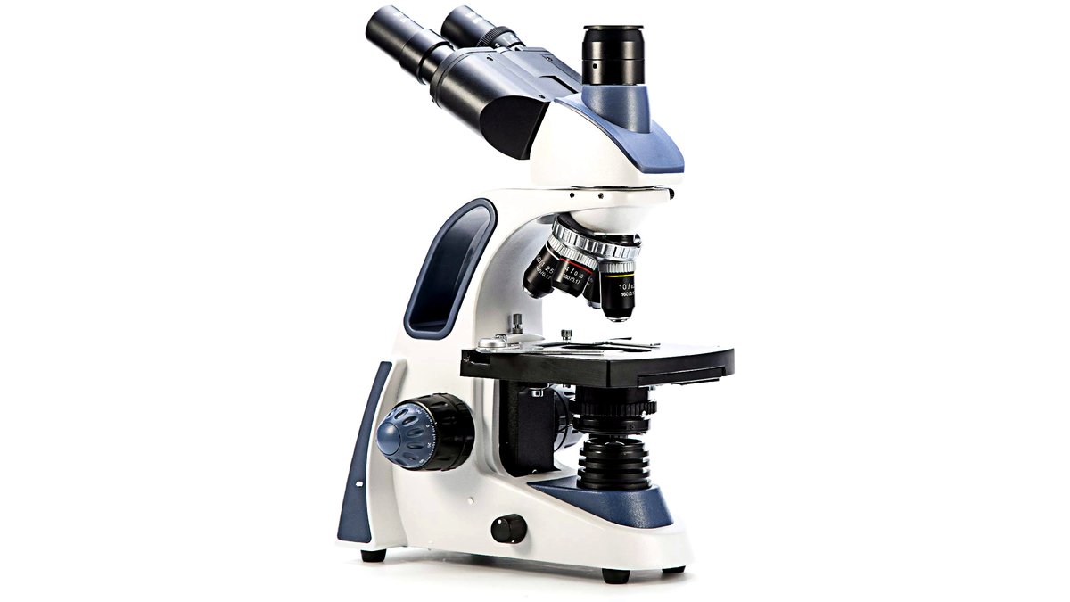
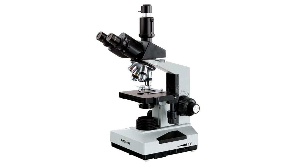
Most current research microscopes support a camera port or trinocular head for attaching a digital camera, which research outlets and instrument makers note has become close to standard expectation for any lab that needs to document, measure, or share images rather than relying on eyepiece observation alone. Camera resolution and sensor type matter for image quality, but just as important is confirming the camera and software are compatible with the specific microscope’s camera port and that the software supports the measurement or analysis tools the lab actually needs, such as basic length and area measurement or more advanced cell-counting routines.
Build quality, stage, and long-term lab use
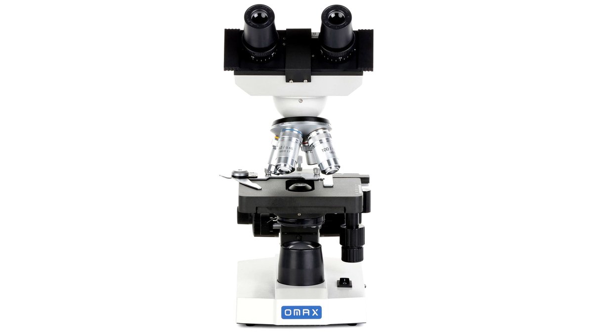
A microscope used daily across many years in a working lab benefits from a mechanical stage (rather than a simpler manual sliding stage) for precise, repeatable specimen positioning, and from a solidly built frame that resists drift and vibration during extended viewing or imaging sessions. Instrument makers generally differentiate their product lines into student/educational, routine clinical or research, and advanced research tiers, with build quality, optical coating quality, and long-term parts and service support scaling up through those tiers.
| Tier | Best for | Key stated traits | Limits |
|---|---|---|---|
| Student/educational compound scope | Teaching labs and basic coursework | Standard brightfield illumination, basic objective set, simpler stage | Lower optical resolution ceiling; usually no phase contrast or fluorescence option |
| Routine research compound scope | Most working biology labs doing cell and tissue observation | Kohler illumination, mechanical stage, often upgradeable to phase contrast | Fluorescence typically not included unless specified |
| Phase contrast research scope | Live cell culture work and unstained specimen observation | Matched phase objectives and condenser, strong contrast on transparent specimens | Adds cost over standard brightfield-only models |
| Fluorescence-capable research scope | Immunofluorescence, tagged-protein, and advanced imaging work | Dedicated fluorescence illuminator, filter cubes matched to fluorophores | Highest cost tier; filter sets must match the specific fluorophores used |
Pick this if…
| If you… | Choose |
|---|---|
| Teach coursework or do basic specimen observation | A student/educational compound microscope |
| Run routine histology, cell counts, or general lab observation | A routine research compound scope with Kohler illumination and a mechanical stage |
| Study live, unstained cells regularly | A phase-contrast-equipped research scope |
| Work with tagged proteins or fluorescent reporters | A fluorescence-capable research scope matched to your specific fluorophores |
| Need to document or measure images digitally | A scope with a trinocular head and camera compatibility confirmed with your software |
Alternatives worth considering
A stereo (dissecting) microscope, which gives a lower-magnification 3D view rather than a thin-section 2D image, is the better tool for dissection, sorting specimens, or working on whole small organisms rather than thin tissue sections or cell monolayers. A digital-only microscope, which replaces the eyepiece entirely with a screen and built-in camera, can suit labs prioritizing easy image capture and sharing over traditional optical viewing, though image quality and resolution on these units vary widely by price tier. For labs needing extremely high-resolution imaging below the diffraction limit of light, a confocal or super-resolution system is a different and far more expensive category entirely, generally reserved for specialized core facilities rather than individual lab purchases.
Frequently Asked Questions
What magnification do I actually need for cell biology work?
Most routine cell observation happens in the 100x to 400x range (10x or 40x objectives with a 10x eyepiece), while examining fine subcellular detail often calls for 1000x using a 100x oil-immersion objective. Buying a scope advertised at extremely high magnification numbers without matching objective and condenser quality rarely produces a usable image at that magnification.
Do I need phase contrast if I mostly work with stained, fixed samples?
Not necessarily. Phase contrast mainly benefits unstained, living, or otherwise low-contrast transparent specimens; stained and fixed tissue sections usually show good contrast under standard brightfield illumination without needing the added phase contrast hardware.
Can I add fluorescence capability to a standard compound scope later?
In some cases yes, if the specific model’s manufacturer offers a compatible add-on fluorescence illuminator and matching objectives, but this isn’t universal across brands and models. It’s generally simpler and more reliable to specify fluorescence capability at the time of purchase if there’s a reasonable chance the lab will need it.
How important is the camera versus the optical system?
The optical system (objectives, condenser, illumination) sets the ceiling for image quality; a camera can only capture what the optics actually resolve, not improve on it. That said, a mismatched or incompatible camera can still bottleneck practical image quality and usability, so checking camera-to-microscope compatibility matters once imaging is part of the workflow.
What maintenance does a research microscope need?
Routine care includes keeping optical surfaces clean with appropriate lens-cleaning materials, protecting the scope from dust with a cover when not in use, and periodically checking illumination alignment, particularly for Kohler-illuminated systems. Fluorescence light sources, especially older mercury or metal-halide bulbs, typically have a stated service life and should be tracked and replaced per the maker’s schedule.
Is a trinocular head worth the extra cost over a standard binocular scope?
If the lab needs to document images, share them for publication, or run any camera-based measurement, yes, since the trinocular port lets a camera run simultaneously with eyepiece viewing. Labs that only ever need direct visual observation without documentation can skip it without losing any imaging capability they’d actually use.